Assistant professor Cliff Stains and his colleagues have detailed the repurposing of protein kinase activity sensors for the direct detection of protein phosphatase activity. Utilizing this approach, the activity of endogenous PTP1B was monitored in response to insulin stimulation as well as in a rat model of nonalcoholic fatty liver disease.
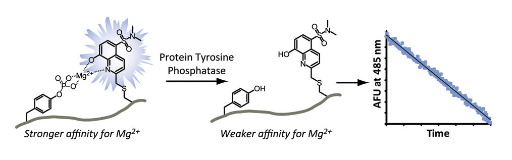
Protein phosphatases have traditionally taken a “backseat” in comparison to relatively more well-studied protein kinases.1 However, a clear role for protein tyrosine phosphatases (PTPs) as signaling nodes in their own right is now emerging. In particular, the PTP known as PTP1B now has a demonstrated role in insulin signaling2 and is implicated in several human disease states including nonalcoholic fatty liver disease (NAFLD).3,4 Thus there is a growing need for methodologies to directly assess endogenous levels of protein phosphatase activity in disease models. To address this issue, the Stains laboratory has recently described an approach to quantify PTP activity from biologically relevant samples. This approach utilizes the phosphorylation-sensitive Sox fluorophore5 to detect phosphate removal in real-time (Fig. 1a). Utilizing this approach, the team was able to construct a sensitive probe, termed PTP1Btide-pS3, capable of detecting a little as 6 pM recombinant PTP1B (Fig. 1b). Leveraging the specificity of immunoaffinity chromatography,6 the researchers were able to directly assess changes in PTP1B activity during insulin stimulation as well as in a rat model of NAFLD (Fig. 1c and d). This collaborative work involved expertise from both the Stains laboratory as well as Edward Harris in the Department of Biochemistry and provides direct evidence for alterations in PTP1B catalytic activity in NAFLD. The team is currently utilizing this approach to assess activity perturbations across the PTP family in various disease states including NAFLD. In the long term, this work lays to groundwork for the potential development of probes capable of functioning in unfractionated samples.
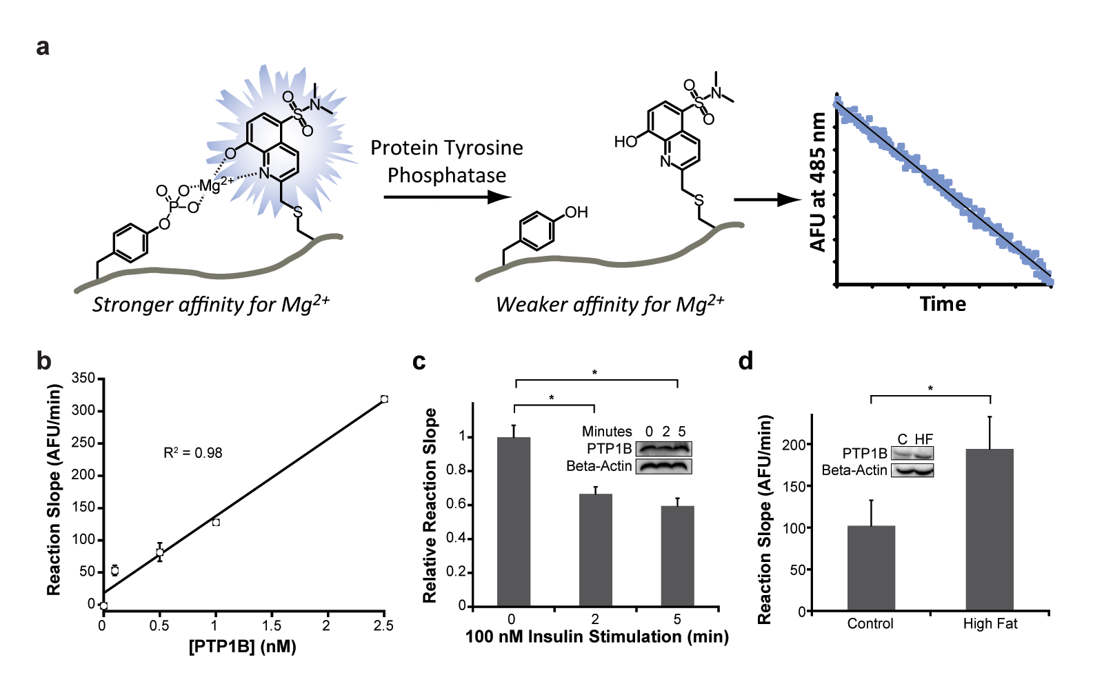
Figure 1. Direct activity sensors for protein phosphatases. (a) Dephosphorylation of a Sox-containing peptide substrate (left) leads to a decrease in fluorescence (middle). The rate of fluorescence decrease is proportional to PTP activity (right) (b) Reaction slopes are proportional to PTP1B concentration, yielding a limit of detection of 6 pM. (c) Analysis of PTP1B activity from HepG2 lysates stimulated for the indicated time period with insulin. Changes in PTP1B catalytic activity are not due to changes in protein concentration (inset) (d) A clear 90% increase in PTP1B activity is observed in high fat (NAFLD) livers from a rat model of NAFLD. This increase in PTP1B activity is partially due to an increase in PTP1B expression (58%) in high fat livers.
The study was published in ACS Chemical Biology and is viewable at https://pubs.acs.org/doi/abs/10.1021/acschembio.5b00506
References:
- Manning, G., Whyte, D.B., Martinez, R., Hunter, T. & Sudarsanam, S. The protein kinase complement of the human genome. Science 298, 1912-1934 (2002).
- Elchebly, M. et al. Increased insulin sensitivity and obesity resistance in mice lacking the protein tyrosine phosphatase-1B gene. Science 283, 1544-1548 (1999).
- Lam, N.T. et al. Leptin resistance following over-expression of protein tyrosine phosphatase 1B in liver. J. Mol. Endocrinol. 36, 163-174 (2006).
- Sanderson, S.O. & Smyrk, T.C. The use of protein tyrosine phosphatase 1B and insulin receptor immunostains to differentiate nonalcoholic from alcoholic steatohepatitis in liver biopsy specimens. Am. J. Clin. Pathol. 123, 503-509 (2005).
- Shults, M.D., Janes, K.A., Lauffenburger, D.A. & Imperiali, B. A multiplexed homogeneous fluorescence-based assay for protein kinase activity in cell lysates. Nat. Methods 2, 277-283 (2005).
- Moser, A.C. & Hage, D.S. Immunoaffinity chromatography: an introduction to applications and recent developments. Bioanalysis 2, 769-790 (2010).